Stroke Localization Part 3- An Easy way to Learn the Brainstem Vascular Syndromes
- Apr 3, 2023
- 4 min read
Want to stimulate the neurologist's' neurons? Give them a stroke localization case, and they will be the happiest people on the world. Solving this riddle is similar to solving a mystery puzzle. It only takes 3 steps.
Step 1: Learning the Neuroanatomy
Step 2: Learn its functions (Patient presents with these symptoms)
Step 3: Match the symptoms with the Neuroanatomy
Anatomically the Central nervous system is divided as the following,
Cerebrum -> Internal Capsule -> Subcortical region -> Midbrain -> Pons -> Medulla -> Spinal cord
A clot can be lodged into any of the vessels supplying the central nervous system. So we need to learn what is inside these parts and its corresponding blood supply.
This article will focus on Brainstem Strokes. Learning the structures at the each level of the brainstem is important. But we all get confused here. Many easy methods, such as The Gate's Rule of 4 published in 2005 (1) and The Rule of 5, Rule of 12, and the Rule of M/S published in 2014 (2) , have been proposed to make it simpler for medical students to follow. Based on which I have consolidated an approach that can be learnt within minutes.
The two main anatomical structures found at the brainstem are the cranial nerve nuclei and the longitudinal tracts. Apart from these, there are many other structures which we will not discuss in this post as it can complicate things. To simply our diagrams, The brainstem can be represented as the following (figure 1)

Let's start by learning about the cranial nerve nuclei.

Look at this diagram (figure 2 ). The cerebral cortex gives out corticobulbar fibres which traverse through the internal capsule to reach the brainstem. This is where 10 out 12 cranial nuclei are present. the 1 and 2 being located above the brainstem.
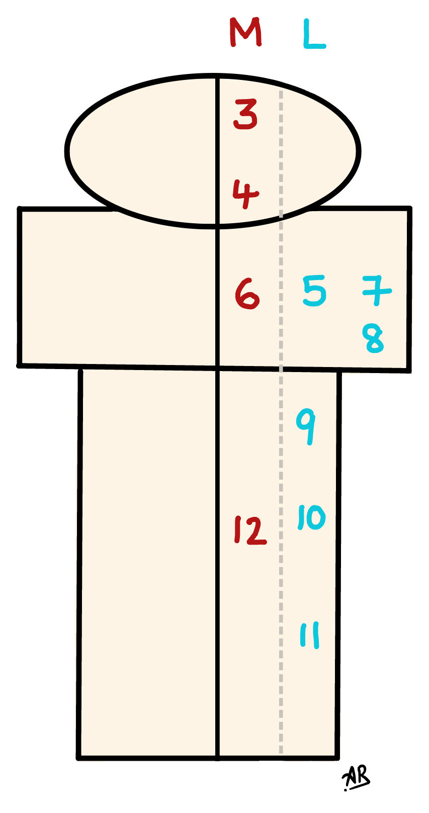
According to rule of 4, the 12 cranial nerve nuclei are evenly distributed as 4 in the midbrain and above, 4 at the pons and 4 located in the medulla. Out of which, all the motor nuclei are to middle (3, 4, 6, 12) and the mixed as well as pure sensory nuclei (5, 7, 8, 9, 10, 11) are to the side. The following diagrams (figure 3, 4) helps us understand this concept.
Tip to remember: Motor = Medial

Next let's learn about the various tracts that traverse the brainstem. Here, we use the M/S rule which is a mnemonic to remember the tracts that run medially and laterally.
The tracts which are medial start with the letter M - Motor pathway (corticospinal tract), Medial lemniscus pathway (DCML) & Medial Longitudinal Fasciculus (MLF). The tracts which are lateral start with the letter S - Sympathetic pathway, Spinothalamic pathway & Spinocerebellar pathway. The following diagrams (figure 5, 6) helps us understand this concept.


Next we need to learn the blood supply of the brainstem. The Anterior spinal artery and Basilar artery supplies the medial portion. The Posterior Inferior cerebellar artery (PICA), Anterior Inferior Cerebellar Artery (AICA) and the Superior Cerebellar Artery (SCA) supply the lateral portion. This is represented diagrammatically in figure 7. A stroke in any of these arteries will result in the corresponding syndrome as given in figure 8.


Hence, the syndromes, its corresponding structures affected and its symptoms can be summarized as given in Table 1.

There are a few focal pontine syndromes that do not follow any rules, due to involvement of certain branches of the basilar artery. If the whole basilar artery is involved at its root level, then it causes locked in Syndrome where pretty much all spinal and bulbar functions are lost except blinking. These are summarised in Table 2.

When it comes to Midbrain syndromes, they do not strictly follow any hard and fast rules. The various named syndromes are identified based on the involvement of branches of Posterior Cerebral Artery (PCA). These are summarised in Table 3.
Tips to Remember:
PCA is the key artery involved in Midbrain syndromes.
Cranial nerve 3 is involved in all the syndromes. The additional structures involved is based on the level and proximity of the vessel that gives rise to unique and specific syndromes.
The midbrain syndromes in the table are arranged in reverse alphabetical order. 'W - Weber' 'N - Nothnagel' 'C - Claude' 'B - Benedikt'. The first and last syndromes involve cerebral peduncles and the middle two syndromes involve the cerebellar peduncle.
The Weber reminds us of a 'Web' - which means it has a lot of tracts. Hence involvement of the corticobulbar as well as corticospinal covering the whole body like a web.
Remember Nothnagel as 'North'nagel and hence the involvement of one and only 'superior' cerebellar peduncle.

Congrats on reaching the end of this post. Just practise matching the symptoms, the vessels involved, and the name of the syndromes.
And That's It!! You have mastered the Brainstem syndromes.
References
Gates P. The rule of 4 of the brainstem: a simplified method for understanding brainstem anatomy and brainstem vascular syndromes for the non-neurologist. Intern Med J. 2005 Apr;35(4):263-6. doi: 10.1111/j.1445-5994.2004.00732.x. PMID: 15836511.
McDeavitt JT, King KC, McDeavitt KR. Learning brainstem anatomy: a mnemonic device. PM R. 2014 Oct;6(10):963-6. doi: 10.1016/j.pmrj.2014.03.013. Epub 2014 Apr 5. PMID: 24713180.

Comments